

How Modern Gastroscopy Equipment Improves Diagnosis in Private Clinics
Last update : 07 Sep 2026
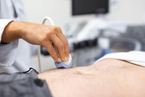
Standard gastroscopies can miss subtle lesions due to outdated lighting and resolution. Advanced private clinics utilize 4K imaging, NBI, and AI-assisted detection to identify early inflammation, ulcers, and Barrett’s esophagus accurately while improving patient comfort with slimmer scope designs.
Key Takeaways
- Diagnostic Precision: Modern gastroscopy technology, including 4K optics, NBI, and AI integration, drastically improves lesion detection and diagnostic accuracy
- Enhanced Comfort: Upgraded, slimmer endoscope designs minimize patient discomfort, reduce the gag reflex, and shorten post-procedure recovery times
- Essential Preparation: Patient preparation and fasting remain critical, as uncleaned gastric mucosa can still obstruct even the most advanced imaging
Choosing where to have an upper GI exam in Canada might feel like a small decision. But it’s only until you realize how much the underlying technology affects what your doctor can actually see during the test.
Two clinics offering the same procedure can produce very different diagnostic results depending on the instruments they use. This blog will explain what modern gastroscopy technology includes, how it changes accuracy and comfort, and what you should look for before booking an appointment yourself.
What “Modern” Gastroscopy Equipment Actually Means
Not all gastroscopy equipment used in Canadian private and public clinics qualifies as advanced. A modern setup is defined less by manufacturing date and more by the following three components:
- The image processor
- The light source
- The endoscope itself
Older instruments rely on halogen or xenon lighting and standard sensors that flatten subtle color differences in the mucosal lining. Modern equipment used in endoscopy implements LED illumination with high-definition or 4K sensors, which show better contrast between healthy and inflamed tissue.
| Component | Traditional Systems | Modern Systems | Diagnostic Impact |
| Light source | Xenon/halogen lamp | LED illumination | More consistent color rendering, longer bulb life, less heat |
| Image sensor | Standard-definition CCD | High-definition/4K CMOS | Sharper detail on small mucosal changes |
| Image processor | Basic video processor | AI-integrated processor | Real-time flagging of suspicious areas |
| Enhancement modes | White light only | NBI, BLI, LCI, i-Scan | Improved contrast for vessels and surface patterns |
| Scope design | Wider, less flexible | Slimmer, high-definition optics | Better patient tolerance, fewer gag-reflex issues |
| Documentation | Static printed images | HD video + still capture, digital storage | Easier specialist review and second opinions |
Table 1: Gastroscopy Equipment Comparison
Clinics investing in this advanced tier of equipment are typically better positioned to catch small lesions, early ulceration, or mucosal changes that basic white-light systems can miss entirely.
How Advanced Endoscopy Equipment Improves Diagnostic Accuracy
The clinical value of advanced gastroscopy equipment shows up in three measurable ways:
- First, image-enhancement technologies such as Narrow Band Imaging (NBI) use specific light wavelengths to increase contrast between blood vessels and surrounding tissue. This makes it easier to spot abnormal vascular patterns linked to early cancers or Barrett’s esophagus.
- Second, higher-resolution optics reduce the chance that a small polyp, ulcer, or inflamed patch is missed because it blends into the surrounding mucosa on a lower-quality screen.
- Third, some current platforms include AI-assisted computer-aided detection, which flags irregular areas in real time as the physician moves the scope, acting as a second set of eyes during the exam.
FACT
Narrow Band Imaging (NBI) enhances surface and vascular detail using specific light wavelengths rather than dye, letting specialists spot suspicious tissue patterns in seconds, a technique now standard on many high-end endoscopy platforms used across Canadian gastroscopy private clinics.
Here’s how some of the gastroscopy technologies work to improve diagnostic accuracy.
| Technology | How It Works | Best For |
| NBI (Narrow Band Imaging) | Filters light to specific wavelengths that highlight blood vessels | Detecting Barrett’s esophagus, early gastric cancer, vascular lesions |
| BLI (Blue Light Imaging) | Combines white light with blue laser for surface contrast | Highlighting mucosal and vascular irregularities |
| LCI (Linked Color Imaging) | Exaggerates subtle red/white color differences | Spotting early inflammation and flat lesions |
| i-Scan | Digital post-processing contrast/surface enhancement, no extra hardware | Chromoendoscopy-style views without dye |
| AI-CAD (Computer-Aided Detection) | Software analyzes the live video feed and flags irregular areas in real time | Reducing missed lesions, standardizing detection rates |
Table 2: Gastroscopy Technology & How They Work
Why This Matters More in a Private Gastroscopy Clinic Setting
Private clinics compete on turnaround time, comfort, and diagnostic confidence. This pushes many clinics to upgrade equipment more often than larger public institutions working within fixed capital budgets.
A well-equipped digestive health clinic can typically complete same-day image review, generate higher-resolution biopsy targeting, and give referring physicians a clearer report package.
For patients weighing a private option against a public wait list, the equipment your choice of clinic uses is one of the few genuinely comparable, verifiable differences between providers, alongside physician experience and accreditation status.
USE CASE
A 52-year-old patient with unexplained iron-deficiency anemia underwent gastroscopy using HD imaging with NBI. The enhanced contrast identified a small bleeding gastric ulcer missed on a prior standard-definition exam, allowing immediate cauterization during the same procedure.
Also read: 10 Tips for Smooth Recovery After Your Colonoscopy or Gastroscopy
Choosing a Digestive Health Clinic with the Right Technology
You might not have direct access to a clinic’s procurement records, but if you ask some practical questions, they can reveal a lot. For starters, before booking your appointment or treatment, make sure you ask the clinic the following questions.
| What to Ask | Why It Matters |
| Is imaging HD or 4K? | Higher resolution reduces missed small lesions |
| Which enhancement modes are available (NBI/BLI/LCI)? | Improves detection of vascular and surface abnormalities |
| Is AI-assisted detection used? | Adds a real-time second layer of lesion flagging |
| How is the endoscope reprocessed/sterilized? | Confirms infection-control standards between patients |
| Is the facility accredited? | Verifies safety and quality benchmarks are independently reviewed |
| How are results documented and shared? | Affects speed and clarity of follow-up care |
Table 3: Things to Ask the Clinic Staff
A transparent digestive health clinic will typically list its imaging technology, accreditation status, and physician credentials openly on its website or share them on request.
USE CASE
A patient with chronic reflux had a gastroscopy using AI-assisted computer-aided detection. The system flagged tissue changes in the lower esophagus, prompting a targeted biopsy that led to early diagnosis of Barrett’s esophagus and a monitoring plan.
Patient Comfort and Safety with Modern Equipment
Newer equipment used in endoscopy also tends to improve the patient experience.
- Slimmer, more flexible scope designs reduce gag reflex and throat discomfort during insertion
- LED light sources generate less heat and allow steadier illumination throughout longer exams
- Many clinics pair this hardware with lighter sedation protocols and shorter recovery-room stays
When comparing your clinic choices, endoscopy equipment quality often correlates with a better overall visit, from prep instructions through discharge.
CAUTION
Advanced imaging improves detection but does not replace preparation. A poorly cleansed stomach or incomplete fasting can obscure the mucosal lining regardless of equipment quality, so following pre-procedure fasting and medication instructions closely remains essential for an accurate result.
Conclusion
The technology working behind a gastroscopy screen has a real and measurable effect on what a physician can find and how comfortable the exam feels. You, as patients, weighing where to book an exam, have every reason to ask about the instruments involved, not just the appointment timing. Essentially, before making a decision about your digestive health and service booking.
Worried About Your Gastroscopy Test Quality? We Use Advanced Gastroscopy Instruments to Ensure Best Quality Results
If you are looking for safe, comfortable, and reliable gastroscopy services in West Island, Laval, or Montreal, Scope MD can help you. Contact us for more information or to book now!
Frequently Asked Questions (FAQs)
1. Does Insurance Or Provincial Health Coverage Pay For Advanced Gastroscopy Technology At Private Clinics?
Basic procedures are often covered, but private facility fees or specialized AI diagnostic add-ons usually require out-of-pocket payment or private insurance.
2. How Long Does A Modern Gastroscopy Procedure Take From Start To Finish?
The actual examination takes 10 to 15 minutes, though total clinic time including preparation and recovery is around 1 to 2 hours.
3. Are There Additional Risks Associated With AI-Assisted Or High-Definition Gastroscopy Devices?
No, advanced imaging relies entirely on optical software, light wavelengths, and processing algorithms, posing no additional physical risk to the patient.
4. How Soon After An Advanced Gastroscopy Can I Return To Normal Daily Activities?
If mild sedation is used, you can resume normal activities the next day, though you must avoid driving for 24 hours.
5. Can Modern Gastroscopy Technology Fully Replace The Need For Physical Tissue Biopsies?
While advanced imaging pinpoints suspicious areas with high precision, physical biopsies remain necessary for definitive microscopic and histological tissue diagnosis.